facs flow cytometry protocol
Load cells into the FACS using using appropriate 5 ml tubes Make sure the instrument is running well by using control samples or any extra cells that you have Replace cell collection tubes and make sure they are sterile if you plan on culturing cells that are collected Run the FACS instrument Notes on this FACSFlow Cytometry Methodology. Collect cells by centrifugation and aspirate supernatant.

Purification Of Micronuclei From Cultured Cells By Flow Cytometry Star Protocols
Indirect flow cytometry FACS protocol General procedure for flow cytometry using a primary antibody and conjugated secondary antibody.
. Protocol Steps Harvest Tissue or Cells. RNA Flow Cytometry Using the Branched DNA Technique. Preparation of cells for flow cytometry.
It also offers an. Obtain desired tissue eg. Leukocytes can be obtained from whole blood and a variety of tissues such as spleen lymph node bone marrow and thymus.
Prepare your cell suspensions for Flow Cytometry Although most flow cytometry experiments involve labeling populations of cells that are relatively abundant the number of cells required will vary depending upon the rarity of your cells. Flex-T Fixed Peptide Tetramer Preparation and Flow Cytometry Staining Protocol. This allows a large number of cells to be analyzed and sorted in a short amount of time.
Cell Preparation Flow Cytometry Protocols I. Ad LabSat Research by Lunaphore is a groundbreaking immunohistochemical staining solution. Resuspend cells in 051 ml 1X PBS.
Proliferation Monitoring by Flow Cytometry webinar from CYTO U. Ad Detect up to 14 colors from 4 lasers with optional imaging and 6 channel violet laser. Preparation of human peripheral blood mononuclear cells.
Intracellular Cytokine Staining Protocol Immunofluorescent Staining of Intracellular Cytokines for Flow Cytometric Analysis. Do not add sodium azide to buffers if you are concerned with recovering cell function eg. How Cytometers Work Basic Operation of a Cell Sorter MACSQuant User.
Flex-T Tetramer Preparation and Flow Cytometry Staining Protocol. 12 To prepare a 10 mM solution of EdU add 4 mL of DMSO Component C or aqueous solution PBS to Component A and mix well. RNA Flow Cytometry webinar from CYTO U.
Compatible with optional autosamplers for walk-away automation and robotic integration. Flow Cytometry Fundamental Principle How FACS Works. Download our brochure now for more information on how LabSat can help your laboratory.
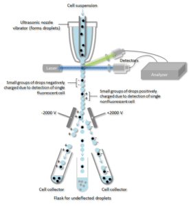
Characterization detection and counting of metal nanoparticles using flow cytometry. FACS is an abbreviation for fluorescence-activated cell sorting which is a flow cytometry technique that further adds a degree of functionality. Protocol Summary of Protocol Preparing Reagents 11 Allow vials to warm to room temperature before opening.
In general researchers will stain between 1 x 10⁵ and 1 x 10⁶ cells per sample. Harvest wash the cells single cell suspension and adjust cell number to a concentration of 1-5106 cellsml in ice cold FACS Buffer PBS 05-1 BSA or 5-10 FBS 01 NaN3 sodium azide. Ideal Shipping Method According To Items Temperature Requirement.
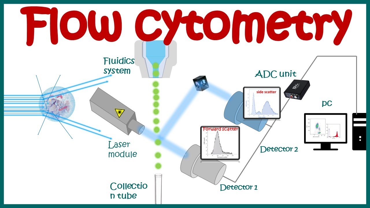
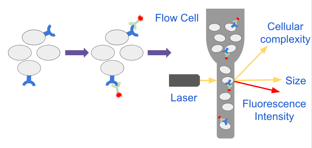
Flow Cytometry is a widely used technique for identifying cell populations as well as measuring cell surface and intracellular molecules. Store the cell suspension immediately at 4C in the dark. Flow cytometry is a popular cell biology technique that utilizes laser-based technology to count sort and profile cells in a heterogeneous fluid mixture.
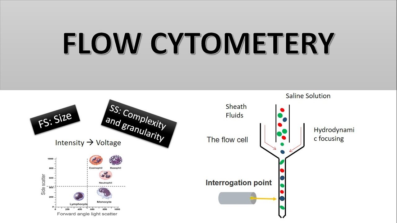
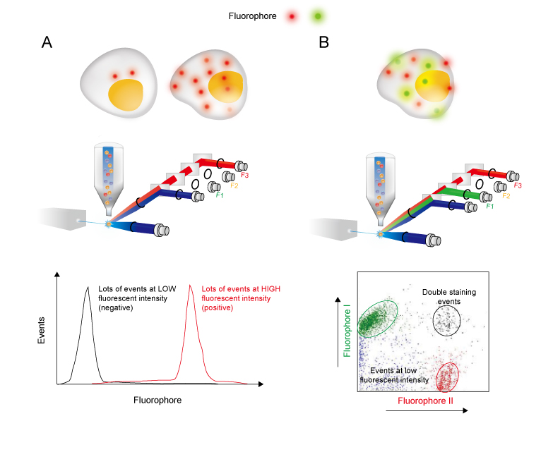
Wash by centrifugation with excess 1X PBS. The properties measured include a particles relative size relative granularity or internal complexity and relative fluorescence intensity. Fix for 15 min at room temperature.
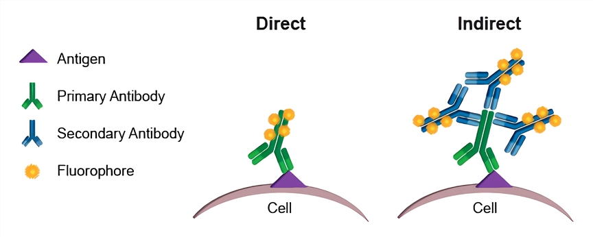
Indirect labelling requires two incubation steps firstly with a primary antibody then with a compatible secondary antibody. Flow cytometry FACS staining protocol Cell surface staining 1. Ad Used To Preserve Cell Surface Epitopes That Have Previously Been Stained.
By utilizing highly specific antibodies labeled with fluorescent conjugates a fluorescent molecule called fluorochrome FACS analysis allows us to simultaneously collect data on and sort a biological sample by a. Cell Surface Flow Cytometry Staining Protocol. Red blood cells RBC present in whole blood or cell preparations are a.
The secondary and not the primary antibody has the fluorescent dye FITC PE Cy5 etc conjugated. Cells flow single file through a flow cytometer and multiple parameters can be collected and measured at a high-speed rate. Ideal Shipping Method According To Items Temperature Requirement.
Get information on stimulation of cells appropriate cultures for generating human mouse and rat cytokine producing cells and describes a protocol for multicolor staining of intracellular cytokines and cell surface antigens. Wash with 2 mL FACS buffer. Whole Blood Staining Protocol for Flow Cytometry Analysis Immune cell stimulation In vitro differentiation of macrophages from monocytes via M-CSF T-cell activation via functional antibodies anti-CD3 and anti-CD28 Immune cell activation for cytokine production via LPS Stimulation of cytokine production in immune cells.
Ad We offer the equipment used for flow cytometry referred to as a flow cytometer. Compatible with optional autosamplers for walk-away automation and robotic integration. Ad We offer the equipment used for flow cytometry referred to as a flow cytometer.
Cell Surface Flow Cytometry Staining of Whole Blood. Place samples in 12 x 75 mm Falcon tubes and analyze by flow cytometry as soon as possible within. Centrifuge and aspirate supernatant.
Ad Used To Preserve Cell Surface Epitopes That Have Previously Been Stained. Resuspend cells with 052 mL FACS buffer. Guide to FACS DiVa pdf.
FACS is an abbreviation for fluorescence-activated cell sorting which is a flow cytometry technique that further adds a degree of. Do not add sodium azide to buffers if you are concerned with recovering cell function eg. Bio-Rad Flow Cytometry Protocols.
Add formaldehyde to obtain a final concentration of 4. By utilizing highly specific antibodies labeled with fluorescent conjugates FACS analysis allows us to simultaneously collect data on and sort a biological sample by a nearly limitless number of different parameters. Ad LabSat Research by Lunaphore is a groundbreaking immunohistochemical staining solution.
Guide to CellQuest Pro pdf. Cytometry in Studies of Programmed Cell Death. Discard supernatant in appropriate waste container.
Ad Detect up to 14 colors from 4 lasers with optional imaging and 6 channel violet laser. Antibody Titration Protocol pdf. Veri-Cells Phospho PBMC MAPKERK.
Cell Preparation Flow Cytometry Protocols Below are protocols for harvesting cells from various sources to obtain healthy cells essential for optimal staining and analysis. This incubation must be done in the dark. Preparation of human peripheral blood mononuclear cells PBMC Leukocytes are the most commonly analyzed cells in flow cytometry.
Wash the cells 3 times by centrifugation at 400 g for 5 min and resuspend them in ice-cold PBS 3 BSA 1 sodium azide. Precision Count Beads Protocol and Applications. Preparation of peritoneal macrophages bone marrow thymus and spleen cells.
General Cell Staining Protocol for Flow Cytometry pdf. Spleen lymph node thymus bone marrow and prepare a single cell suspension in Cell Staining Buffer BioLegend Cat. FACS is an abbreviation for fluorescence-activated single cell sorting which is a flow cytometry technique that further adds a degree of functionality.
Download our brochure now for more information on how LabSat can help your laboratory. If using in vitro stimulated cells simply resuspend previously activated cultures in Cell Staining Buffer and proceed to Step 2. Resuspend cells in 05-1 ml 1X PBS.
After use store any remaining stock solution at 20C. Incubate for at least 20-30 min at room temperature of 4C. Flow cytometry is a technology that simultaneously measures and then analyzes multiple physical characteristics of single particles usually cells as they flow in a fluid stream through a beam of light.
Secondary antibodies used for FACS are typically Fab2 fragment antibodies conjugated to fluorochromes like FITC or R-Phycoerythrin. Flow cytometry FACS staining protocol Cell surface staining Harvest wash the cells single cell suspension and adjust cell number to a concentration of 1-5x10⁶ cellsml in ice cold FACS Buffer PBS 05-1 BSA or 5-10 FBS 01 NaN3 sodium azide.

Flow Cytometry Based Protocols For Human Blood Marrow Immunophenotyping With Minimal Sample Perturbation Star Protocols
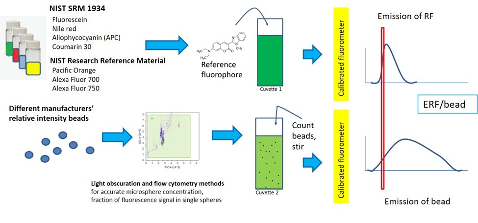
Quantitative Flow Cytometry Measurements Nist
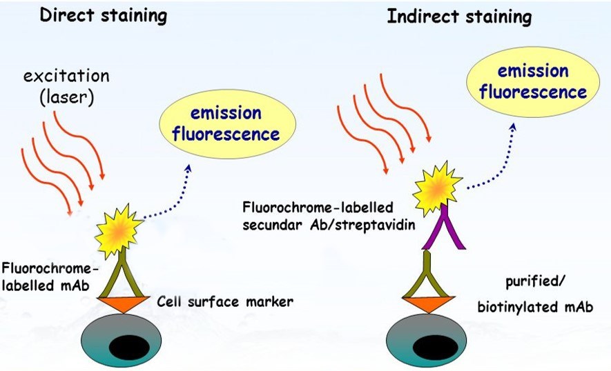
Direct Staining Flow Cytometry Creative Biolabs

Flow Cytometry Illustrated Assay Novus Biologicals

Accelerating Drug Discovery And Development With Flow Cytometry
Flow Cytometry Basic Principles What The Use Of Flow Cytometry Cell Sorting By Facs Youtube
The Principle Of Flow Cytometry And Facs 2 Facs Fluorescence Activated Cell Sorting Youtube

Flow Cytometry Services Promab
Analyzing Single Cells With Flow Cytometry

Flow Cytometry Creative Biolabs

Flow Cytometry Protocols

Flow Cytometry Introduction Abcam

Flow Cytometry Facs Protocols Sino Biological
How Does Flow Cytometry Work Nanocellect
Flow Cytometry Creative Biolabs
Flow Cytometry Guide Creative Diagnostics

In The Protocol Developed By Bernhard Fuchs S Team Bacterial Groups Are Enriched In Three Steps 1 In Situ Hybridization Postdoctoral Researcher Microbiology

Flow Cytometry Sample Preparation Proteintech Group

Optimized Flow Cytometric Protocol For The Detection Of Functional Subsets Of Low Frequency Antigen Specific Cd4 And Cd8 T Cells Sciencedirect